The human eye is a complex and fascinating organ that enables us to perceive and interpret the world around us.
The human eye is a complex and fascinating
organ that enables us to perceive and interpret the world around us. It is a
remarkable example of evolutionary engineering, with various structures working
together to convert light into electrical signals that our brain can
understand. In this article, we will explore the anatomy and main functions of
the eye, making it easy to understand
how this incredible organ works.
The External Eye
The external eye consists of several
visible structures that play crucial roles in protecting the eye and
facilitating vision.
Eyelids are movablefolds of skin that protect the eye by
blinking and spreading tears. Extraocular muscles although not visible
outside are those muscles that are responsible for the eyes to move
synchronously in different directions. Eye
movements help us to scan the visual environment and track objects. There are
several types of eye movements, including saccades, pursuit movements,
and vergence movements.Squint, also known as strabismus, is a
condition where the eyes are misaligned. Squint can be caused by a variety of
factors, including muscle imbalance, refractive errors, and neurological
disorders. Treatment for squint depends on the underlying cause and may include
glasses, patching, or surgery.
The tear film is a thin layer of tears that covers the surface of the eye,
playing a crucial role in maintaining eye health and vision. The tear film helps to lubricate the eye,
reduce friction, and protect against infection. A healthy tear film is
essential for clear vision and comfortable eyes.
Disruption in the tear film leads to
symptoms of dry eye which are irritation, grittiness, redness and
sometimes decreased vision. The commonest mode of treatment is by using lubricating
eye drops.
The cornea is a transparent,
dome-shaped surface at the front of the eye, allowing light to enter. Corneal
transplantation is a surgical procedure where a damaged or diseased cornea
is replaced with a healthy donor cornea. Corneal transplantation is indicated
for conditions such as keratoconus, corneal scarring, and inadequate corneal integrity. Eye
donation is when a deceased person’s cornea is removed and transplanted to
patients who have an opaque cornea.
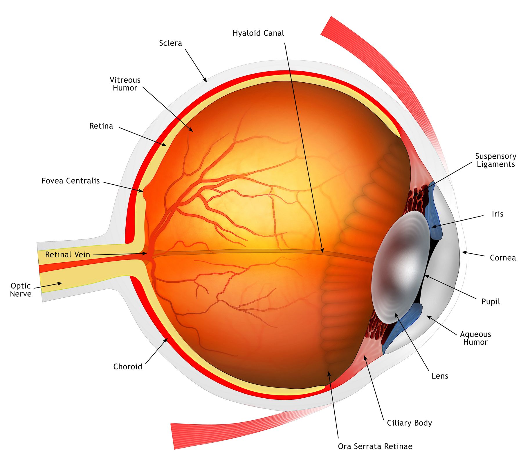
The colored part of the eye, controlling
the amount of light that enters by adjusting the size of the pupil is called
the iris. It contains the pupil which is an opening in the center
of the iris, regulating light entry into the eye. The sclera is the white,
outer layer of the eye, providing protection and structure.
The
sclera is the white portion of the eye surrounding the cornea. It is
rigid and helps in maintaining the shape and integrity of the eyeball. It
extends from the cornea all the way backwards till the optic nerve.In fact the
optic nerve leaves the eyeball to the brain through an opening in the sclera.
Inflammation of the sclera is called scleritis.
The Internal Eye
The internal eye consists of several
structures that work together to focus light and convert it into electrical
signals. The lens is a clear,
flexible structure behind the iris, changing shape to focus light on the
retina. As we age the lens is unable to focus for near object as well, a
condition called aspresbyopia in which reading glasses are
required to read and see objects up close. Cataract is
a condition when there is clouding of the lens, affecting vision. Cataract
surgery by phacoemusification is the commonest ocular surgery
performed worldwide. The natural lens is
removed and replaced with an artificial intraocular lens (IOL) thereby
restoring vision. The choroidis a layer of blood vessels between the sclera and
retina, supplying the retina with oxygen and nutrients. It is the continuity of
the iris at the back of the eye and is the most vascular part of the eye. The
choroid plays a vital role in maintaining the health and function of the
retina. Inflammation of this middle layer can cause iritis if it affects
the front portion and choroiditis if the back portion of this layer is
involved. The vitreous is the gel like
substance that fills the back of the eye. It is a transparent structure and is
important in maintaining the shape of the eyeball and providing nutrients to
the lens. Syneresis is a condition when the vitreous liquifies and collapses due to age. It
separates from the retina which is called posterior vitreous detachment. Anomalous posterior vitreous detachment can lead to various conditions like retinal
tears, vitreous haemorrhage, retinal detachment, vitreomacular
traction and macular hole.
The retina is the innermost layer of the eye, containing specialized light-sensitive cells (photoreceptors) that convert light into electrical signals. The macula is a specialized region at the center of the retina, responsible for central vision and fine detail. Age related macular degeneration is a condition seen in elderly patients when there is damage to the macula causing thinning of the macula, bleeding or fluid leakage into the macular area.
Th optic nerve carries electrical signals from the retina to the brain, enabling us to interpret visual information. It also the portion that gets damaged when the pressure inside the eye (intraocular pressure) is high as in glaucoma. The eye works by focusing light onto the
retina, where photoreceptors convert it into electrical signals. These signals
are then transmitted to the brain, which interprets them as visual information. 1. Light Entry: Light enters the eye
through the cornea and pupil. 2. Focusing: The lens changes shape to
focus light on the retina. 3. Signal Conversion: Photoreceptors in the
retina convert light into electrical signals. 4. Signal Transmission: The optic nerve
transmits electrical signals to the brain. 5. Interpretation: The brain interprets
electrical signals as visual information, enabling us to see and understand the
world. Common Eye Conditions While the eye is a remarkable organ, it can
be susceptible to various conditions that affect vision. Some common eye
conditions include: 1. Myopia (Nearsightedness):
Difficulty seeing distant objects clearly. 2. Hyperopia (Farsightedness):
Difficulty seeing close objects clearly. 3. Astigmatism: Blurred vision due
to irregular corneal shape. 4. Cataracts: Clouding of the lens,
affecting vision. 5.Glaucoma: Optic nerve damage due
to increased intraocular pressure. 6. Age-related Macular Degeneration:
Gradual loss of central vision due to aging. Conclusion
The human eye is a complex and fascinating
organ that enables us to perceive and interpret the world around us.
Understanding its anatomy and functions can help us appreciate the importance
of eye care and vision health. By taking care of our eyes and seeking regular
eye exams, we can maintain good vision and enjoy the world around us.