- Home
- About Us
- General Information
- Eye Speciality
- LASIK – Refractive Surgery
- General Ophthalmology and Cataract
- Cornea
- Treatment for Hereditary Cornea Disorders
- Treatment for Corneal Infections
- Treatment for Allergic Eye Diseases
- Treatment for Keratoconus
- Treatment for Severe Dry Eye
- Treatment for Blepharitis
- Treatment for Chemical Injuries
- Treatment for Refractive Errors
- Keratoplasty
- Corneo Scleral Tear Repair
- Penetrating Keratoplasty
- Refractive Corneal Surgeries
- Vitreo Retina
- Medical Retina
- Glaucoma Treatment
- Strabismus (squint) & Paediatric Ophthalmology
- Orbit and Oculoplasty
- Neuro Ophthalmology
- UVEA
- Support Services
- Blogs
- Training and Education
- Testimonials
- Gallery
- Contact Us


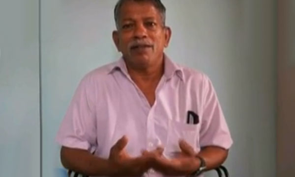
 I am Nimitha, before Lasik, I am very difficult to see and difficult to handle contact lens and specs. After the lasik treatment I am very relaxed.