
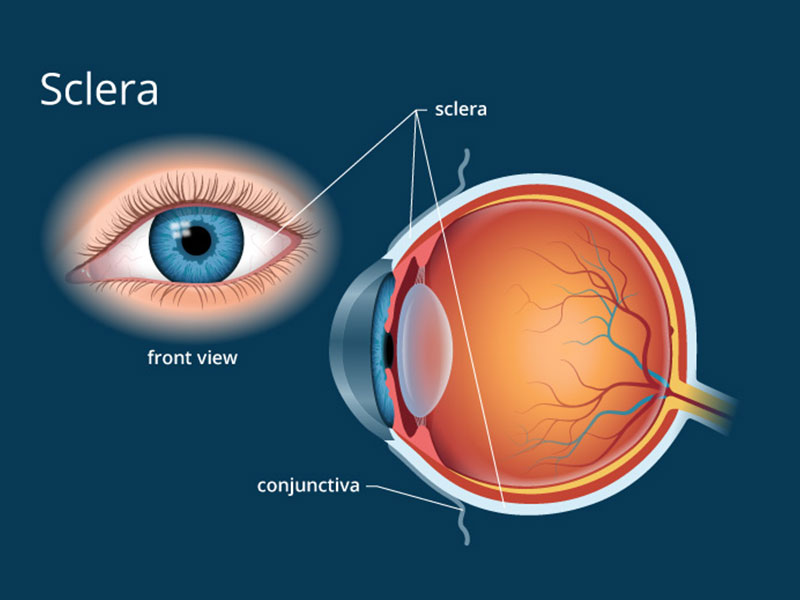
Sclera is a part of the eye & generally known as the “white.” It will form a supporting wall of the eyeball, & is continuous with the clear cornea. In humans, the entire sclera is white contrast with coloured iris, but in the other mammals the visible part of the sclera will equivalent to the colour of the iris, by that white part won’t generally seen. In the embryo development, the sclera is derived from the neural crest. In children, it is thinner & shows some of the underlying pigment, which will appear slightly blue. In elder persons, the fatty deposits on the sclera will make it appear as slightly yellow.
The sclera is covered by conjunctiva, a clear mucus membrane which help to lubricate the eye. It is thickest in the area around the optic nerve.
The sclera is made up of three divisions:
There are a number of abnormalities which are related with the sclera. Some are genetically related abnormalities that may includes:
Acquired abnormalities of sclera which may includes:


 I am Nimitha, before Lasik, I am very difficult to see and difficult to handle contact lens and specs. After the lasik treatment I am very relaxed.
I am Suja Eldho, from London, The brilliant Service has been given to us from Dr. Tony Fernandez Eye Hospital. The staffs have been incredibily
My name is Chanchal Gibson, 45 yrs old , I was having very bad power of Sph -9.00 in right eye and Sph -7.50 in left eye. It was very difficult